Моделювання взаємодії С60 фулерену з білковими мішенями SARS-CoV-2
Анотація
Актуальність: Віднайдення ефективних лікувальних засобів проти небезпечної хвороби, спричиненої SARS-CoV-2, є важливим напрямом біомедичних досліджень. Методи молекулярного докінгу та молекулярної динаміки є ключовими інструментами сучасної фармацевтичної науки, забезпечують швидкий пошук і оптимізацію противірусних сполук, дозволяють прогнозувати їхню ефективність та адаптувати терапію до нових штамів SARS-CoV-2. Фулерен C60 привертає значну увагу як перспективний наноматеріал у боротьбі з SARS-CoV-2 завдяки своїй здатності утворювати стабільні комплекси з ключовими вірусними білками, такими як головна протеаза (3CLpro) та РНК-залежна РНК-полімераза (RdRp). Молекулярні моделювання та біофізичні дослідження показали, що C60 може проникати крізь ліпідну оболонку вірусу та блокувати функціональну активність його білків, що відкриває можливості для створення нових противірусних препаратів. Враховуючи постійні мутації SARS-CoV-2 та обмеженість існуючих терапевтичних засобів, дослідження фулерену C60 як потенційного інгібітора є актуальним напрямом нанотехнології для розробки інноваційних стратегій лікування COVID-19.
Мета роботи полягала в оцінці in silico здатності С60 фулерену взаємодіяти з білковими мішенями 3CLpro (3-Chymotrypsin-Like protease) і RdRp (RNA-dependent RNA polymerase) коронавірусу SARS-CoV-2 і, таким чином, цілеспрямовано їх блокувати, пригнічуючи функціональну активність SARS-CoV-2.
Методи: Структурні дані білків 3CLpro та RdRp коронавірусу SARS-CoV-2 було отримано з Protein Data Bank, а геометрію С₆₀ фулерену згенеровано за допомогою онлайн-сервера SwissParam. Взаємодії між С₆₀ фулереном і досліджуваними білками моделювали за допомогою алгоритму системного молекулярного докінгу (sdock+). Потенційні сайти зв’язування визначали за допомогою програмного пакета Caver. Молекулярно-динамічні розрахунки проводили у програмному середовищі Gromacs 2020. Енергетичну мінімізацію потенційних комплексів «С₆₀ фулерен – білок» виконували з використанням пакета g_mmpbsa.
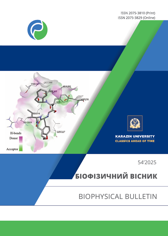
Результати: Встановлено можливий механізм зв’язування С₆₀ фулерену з білковими мішенями 3CLpro та RdRp коронавірусу SARS-CoV-2. Дані молекулярного докінгу та молекулярної динаміки демонструють, що С₆₀ фулерен формує стабільні комплекси з цими білками, що може призводити до пригнічення їх функціональної активності.
Висновки: Показано, що С₆₀ фулерен здатний утворювати стабільні комплекси з білками 3CLpro і RdRp SARS-CoV-2, що потенційно знижує їхню активність і, відповідно, може впливати на загальну активність коронавірусу.
Завантаження
Посилання
Ochani RK, Asad A, Yasmin F, Shaikh S, Khalid H, Batra S, et al. COVID-19 pandemic: from origins to outcomes. A comprehensive review of viral pathogenesis, clinical manifestations, diagnostic evaluation, and management. Infez Med. 2021;29(1):20–36. Available from: https://www.infezmed.it/media/journal/Vol_29_1_2021_3.pdf
Lai A, Bergna A, Ventura CD, Zehender G. Genomic epidemiology and phylogenetics applied to the study of SARS-CoV-2 pandemic. New Microbiol. 2023;46(1):1–8. Available from: https://www.newmicrobiologica.org/PUB/allegati_pdf/2023/1/1.pdf
Rahman S, Hossain MJ, Nahar Z, Shahriar M, Bhuiyan MA, Islam MR. Emerging SARS-CoV-2 variants and subvariants: challenges and opportunities in the context of COVID-19 pandemic. Environ Health Insights. 2022;16:11786302221129396. https://doi.org/10.1177/11786302221129396
Nazmunnahar, Ahmed I, Islam MR. Risk evaluation and mitigation strategies for newly detected SARS-CoV-2 Omicron BF.7 subvariant: a brief report. Health Sci Rep. 2023;6(3):e1127. https://doi.org/10.1002/hsr2.1127
Woo PCY, de Groot RJ, Haagmans B, Lau SKP, Neuman BW, Perlman S, et al. ICTV Virus Taxonomy Profile: Coronaviridae 2023. J Gen Virol. 2023; 104(4):001843. https://doi.org/10.1099/jgv.0.001843
Amorim VMF, Soares EP, Ferrari ASA, Merighi DGS, de Souza RF, Guzzo CR, et al. 3-Chymotrypsin-like Protease (3CLpro) of SARS-CoV-2: Validation as a Molecular Target, Proposal of a Novel Catalytic Mechanism, and Inhibitors in Preclinical and Clinical Trials. Viruses. 2024;16(6):844. https://doi.org/10.3390/v16060844
Hillen HC, Kokic G, Farnung L, Dienemann C, Tegunov D, Cramer P. Structure of replicating SARS-CoV-2 polymerase. Nature. 2020;584:154–6. https://doi.org/10.1038/s41586-020-2368-8
Lee J, Kenward C, Worral1 LJ, Vuckovic M, Gentile F, Ton A-T, et al. X-ray crystallographic characterization of the SARS-CoV-2 main protease polyprotein cleavage sites essential for viral processing and maturation. Nat Commun. 2022;13:5196. https://doi.org/10.1038/s41467-022-32854-4
Kirchdoerfer RN, Ward AB. Structure of the SARS-CoV nsp12 polymerase bound to nsp7 and nsp8 co-factors. Nat Commun. 2019;10:2342. https://doi.org/10.1038/s41467-019-10280-3
Nikaeen G, Abbaszadeh S, Yousefinejad S. Application of nanomaterials in treatment, anti-infection and detection of coronaviruses. Nanomedicine (Lond). 2020;15(15):1501–12. https://doi.org/10.2217/nnm-2020-0117
Innocenzi P, Stagi L. Carbon-based antiviral nanomaterials: graphene, C-dots, and fullerenes. A perspective. Chem Sci. 2020;11(26):6606–22. https://doi.org/10.1039/D0SC02658A
Dechant PP, Wardman J, Keef T, Twarock R. Viruses and fullerenes — symmetry as a common thread? Acta Crystallogr A Found Adv. 2014;70(2):162–7. https://doi.org/10.1107/S2053273313034220
Ritter U, Prylutskyy YuI, Evstigneev MP, Davidenko NA, Cherepanov VV, Senenko AI, et al. Structural features of highly stable reproducible C60 fullerene aqueous colloid solution probed by various techniques. Fullerenes Nanotubes Carbon Nanostruct. 2015;23(6):530–4. https://doi.org/10.1080/1536383X.2013.870900
Halenova T, Raksha N, Savchuk O, Ostapchenko L, Prylutskyy YuI, Ritter U, et al. Evaluation of the biocompatibility of water-soluble pristine C60 fullerenes in rabbit. BioNanoSci. 2020;10:721–30. https://doi.org/10.1007/s12668-020-00762-w
Goodarzi S, Da Ros T, Conde J, Sefat F, Mozafari M. Fullerene: biomedical engineers get to revisit an old friend. Mater Today. 2017;20(8):460–80. https://doi.org/10.1016/j.mattod.2017.03.017
Moussa F. Fullerene and derivatives for biomedical applications. Nanobiomater. 2018:113–36. https://doi.org/10.1016/B978-0-08-100716-7.00005-2
Sijbesma R, Srdanov G, Wudl F, Castoro JA, Wilkins C, Friedman CH, et al. Synthesis of a fullerene derivative for the inhibition of HIV enzymes. J Am Chem Soc. 1993;115(15):6510–12. https://doi.org/10.1021/ja00068a006
Marchesan S, Da Ros T, Spalluto G, Balzarini J, Prato M. Anti-HIV properties of cationic fullerene derivatives. Bioorg Med Chem Lett. 2005;15(15):3615–18. https://doi.org/10.1016/j.bmcl.2005.05.069
Kataoka H, Ohe T, Takahashi K, Nakamura S, Mashino T. Novel fullerene derivatives as dual inhibitors of Hepatitis C virus NS5B polymerase and NS3/4A protease. Bioorg Med Chem Lett. 2016;26(19):4565–7. https://doi.org/10.1016/j.bmcl.2016.08.086
Hurmach V, Karaushu V, Klestova Z, Berest V, Prylutskyy Y. C60 fullerene nanoparticles permeability through the model lipid envelope of coronavirus and their anticoronavirus effect in the in ovo system. Biophysical Bulletin. 2023;(50):17–24. https://doi.org/10.26565/2075-3810-2023-50-02
Hurmach V, Karaushu V, Klestova Z, Berest V, Prylutskyy Y. Anticoronavirus activity of C60 fullerene: in silico and in vitro screening. Biophysical Bulletin. 2025;(53):60–71. https://doi.org/10.26565/2075-3810-2025-53-05
RCSB Protein Data Bank [Internet]. The Research Collaboratory for Structural Bioinformatics Protein Data Bank [cited 2024 Nov 5]. Available from: https://www.rcsb.org/
Zoete V, Cuendet MA, Grosdidier A. SwissParam: a fast force field generation tool for small organic molecules. J Comput Chem. 2011;32(11):2359–68. https://doi.org/10.1002/jcc.21816
McMartin C, Bohacek RS. QXP: powerful, rapid computer algorithms for structure-based drug design. J Comput Aided Mol Des. 1997;11(4):333–44. https://doi.org/10.1023/А:1007907728892
Warren GL, Andrews CW, Capelli A-M, Clarke B, LaLonde J, Lambert MH, et al. A critical assessment of docking programs and scoring functions. J Med Chem. 2006;49(20):5912–31. https://doi.org/10.1021/jm050362n
Abraham MJ, Murtola T, Schulz R, Páll S, Smith JC, Hess B, et al. GROMACS: High performance molecular simulations through multi-level parallelism from laptops to supercomputers. SoftwareX. 2015;1–2:19–25. https://doi.org/10.1016/j.softx.2015.06.001
Huang J, MacKerell Jr AD. CHARMM36 all-atom additive protein force field: validation based on comparison to NMR data. J Comput Chem. 2013;34(25):2135–45. https://doi.org/10.1002/jcc.23354
Jurcik A, Bednar D, Byska J, Marques SM, Furmanova K, Daniel L, et al. CAVER Analyst 2.0: Analysis and Visualization of Channels and Tunnels in Protein Structures and Molecular Dynamics Trajectories. Bioinformatics. 2018;34(20):3586–8. https://doi.org/10.1093/bioinformatics/bty386
Kumari R. g_mmpbsa [Internet]. Available from: https://rashmikumari.github.io/g_mmpbsa/
Wua C, Liu Y, Yang Y. Analysis of therapeutic targets for SARS-CoV-2 and discovery of potential drugs by computational methods. Acta Pharm Sin B. 2020;10:766–88. https://doi.org/10.1016/j.apsb.2020.02.008
Prylutska SV, Grebinyk AG, Lynchak OV, Byelinska IV, Cherepanov VV, Tauscher E, et al. In vitro and in vivo toxicity of pristine C60 fullerene aqueous colloid solution. Fullerenes Nanotubes Carbon Nanostruct. 2019;27(9):715–28. https://doi.org/10.1080/1536383X.2019.1634055
Авторське право (c) 2025 В. В. Гурмач , В. Р. Караушу, З. С. Клестова, В. П. Берест, Ю. І. Прилуцький

Цю роботу ліцензовано за Міжнародня ліцензія Creative Commons Attribution 4.0.
Автори, які публікуються у цьому журналі, погоджуються з наступними умовами:
- Автори залишають за собою право на авторство своєї роботи та передають журналу право першої публікації цієї роботи на умовах ліцензії Creative Commons Attribution License, котра дозволяє іншим особам вільно розповсюджувати опубліковану роботу з обов'язковим посиланням на авторів оригінальної роботи та першу публікацію роботи у цьому журналі.
- Автори мають право укладати самостійні додаткові угоди щодо неексклюзивного розповсюдження роботи у тому вигляді, в якому вона була опублікована цим журналом (наприклад, розміщувати роботу в електронному сховищі установи або публікувати у складі монографії), за умови збереження посилання на першу публікацію роботи у цьому журналі.
- Політика журналу дозволяє і заохочує розміщення авторами в мережі Інтернет (наприклад, у сховищах установ або на особистих веб-сайтах) рукопису роботи, як до подання цього рукопису до редакції, так і під час його редакційного опрацювання, оскільки це сприяє виникненню продуктивної наукової дискусії та позитивно позначається на оперативності та динаміці цитування опублікованої роботи (див. The Effect of Open Access).


