Modeling the interaction of C60 fullerene with SARS-CoV-2 protein targets
Abstract
Background: The discovery of effective therapeutics against the dangerous disease caused by SARS-CoV-2 is an important direction of biomedical research. Molecular docking and molecular dynamics methods are key tools of modern pharmaceutical science, providing rapid search and optimization of antiviral compounds, allowing to predict their effectiveness and adapt therapy to new strains of SARS-CoV-2. Fullerene C60 attracts considerable attention as a promising nanomaterial in the fight against SARS-CoV-2 due to its ability to form stable complexes with key viral proteins, such as the main protease (3CLpro) and RNA-dependent RNA polymerase (RdRp). Molecular modeling and biophysical studies have shown that C60 can penetrate the lipid envelope of the virus and block the functional activity of its proteins, which opens up opportunities for the creation of new antiviral drugs. Given the constant mutations of SARS-CoV-2 and the limitations of existing therapeutics, the study of C60 fullerene as a potential inhibitor is a relevant direction of nanotechnology for the development of innovative strategies for the treatment of COVID-19.
Aim of the work was to assess in silico the ability of C60 fullerene to interact with the protein targets 3CLpro (3-Chymotrypsin-Like protease) and RdRp (RNA-dependent RNA polymerase) of the SARS-CoV-2 coronavirus and, thus, to specifically block them, inhibiting the functional activity of SARS-CoV-2.
Methods: Structural data of the 3CLpro and RdRp proteins of the SARS-CoV-2 coronavirus were obtained from the Protein Data Bank, and the geometry of C60 fullerene was generated using the online server SwissParam. Interactions between С60 fullerene and the studied proteins were modeled using the system molecular docking algorithm (sdock+). Potential binding sites were determined using the Caver software package. Molecular dynamics calculations were performed in the Gromacs 2020 software environment. Energy minimization of potential C60 fullerene — protein complexes was performed using the g_mmpbsa software.
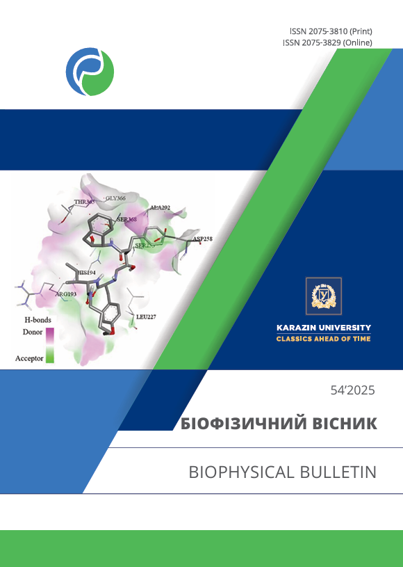
Results: Putative mechanism of binding of C60 fullerene to the protein targets 3CLpro and RdRp of the SARS-CoV-2 coronavirus was established. Molecular docking and molecular dynamics data demonstrate that C60 fullerene forms stable complexes with these proteins, which can lead to inhibition of their functional activity.
Conclusions: It is shown that C60 fullerene is able to form stable complexes with the 3CLpro and RdRp proteins of SARS-CoV-2, which potentially reduces their activity and, accordingly, can affect the overall activity of the coronavirus.
Downloads
References
Ochani RK, Asad A, Yasmin F, Shaikh S, Khalid H, Batra S, et al. COVID-19 pandemic: from origins to outcomes. A comprehensive review of viral pathogenesis, clinical manifestations, diagnostic evaluation, and management. Infez Med. 2021;29(1):20–36. Available from: https://www.infezmed.it/media/journal/Vol_29_1_2021_3.pdf
Lai A, Bergna A, Ventura CD, Zehender G. Genomic epidemiology and phylogenetics applied to the study of SARS-CoV-2 pandemic. New Microbiol. 2023;46(1):1–8. Available from: https://www.newmicrobiologica.org/PUB/allegati_pdf/2023/1/1.pdf
Rahman S, Hossain MJ, Nahar Z, Shahriar M, Bhuiyan MA, Islam MR. Emerging SARS-CoV-2 variants and subvariants: challenges and opportunities in the context of COVID-19 pandemic. Environ Health Insights. 2022;16:11786302221129396. https://doi.org/10.1177/11786302221129396
Nazmunnahar, Ahmed I, Islam MR. Risk evaluation and mitigation strategies for newly detected SARS-CoV-2 Omicron BF.7 subvariant: a brief report. Health Sci Rep. 2023;6(3):e1127. https://doi.org/10.1002/hsr2.1127
Woo PCY, de Groot RJ, Haagmans B, Lau SKP, Neuman BW, Perlman S, et al. ICTV Virus Taxonomy Profile: Coronaviridae 2023. J Gen Virol. 2023; 104(4):001843. https://doi.org/10.1099/jgv.0.001843
Amorim VMF, Soares EP, Ferrari ASA, Merighi DGS, de Souza RF, Guzzo CR, et al. 3-Chymotrypsin-like Protease (3CLpro) of SARS-CoV-2: Validation as a Molecular Target, Proposal of a Novel Catalytic Mechanism, and Inhibitors in Preclinical and Clinical Trials. Viruses. 2024;16(6):844. https://doi.org/10.3390/v16060844
Hillen HC, Kokic G, Farnung L, Dienemann C, Tegunov D, Cramer P. Structure of replicating SARS-CoV-2 polymerase. Nature. 2020;584:154–6. https://doi.org/10.1038/s41586-020-2368-8
Lee J, Kenward C, Worral1 LJ, Vuckovic M, Gentile F, Ton A-T, et al. X-ray crystallographic characterization of the SARS-CoV-2 main protease polyprotein cleavage sites essential for viral processing and maturation. Nat Commun. 2022;13:5196. https://doi.org/10.1038/s41467-022-32854-4
Kirchdoerfer RN, Ward AB. Structure of the SARS-CoV nsp12 polymerase bound to nsp7 and nsp8 co-factors. Nat Commun. 2019;10:2342. https://doi.org/10.1038/s41467-019-10280-3
Nikaeen G, Abbaszadeh S, Yousefinejad S. Application of nanomaterials in treatment, anti-infection and detection of coronaviruses. Nanomedicine (Lond). 2020;15(15):1501–12. https://doi.org/10.2217/nnm-2020-0117
Innocenzi P, Stagi L. Carbon-based antiviral nanomaterials: graphene, C-dots, and fullerenes. A perspective. Chem Sci. 2020;11(26):6606–22. https://doi.org/10.1039/D0SC02658A
Dechant PP, Wardman J, Keef T, Twarock R. Viruses and fullerenes — symmetry as a common thread? Acta Crystallogr A Found Adv. 2014;70(2):162–7. https://doi.org/10.1107/S2053273313034220
Ritter U, Prylutskyy YuI, Evstigneev MP, Davidenko NA, Cherepanov VV, Senenko AI, et al. Structural features of highly stable reproducible C60 fullerene aqueous colloid solution probed by various techniques. Fullerenes Nanotubes Carbon Nanostruct. 2015;23(6):530–4. https://doi.org/10.1080/1536383X.2013.870900
Halenova T, Raksha N, Savchuk O, Ostapchenko L, Prylutskyy YuI, Ritter U, et al. Evaluation of the biocompatibility of water-soluble pristine C60 fullerenes in rabbit. BioNanoSci. 2020;10:721–30. https://doi.org/10.1007/s12668-020-00762-w
Goodarzi S, Da Ros T, Conde J, Sefat F, Mozafari M. Fullerene: biomedical engineers get to revisit an old friend. Mater Today. 2017;20(8):460–80. https://doi.org/10.1016/j.mattod.2017.03.017
Moussa F. Fullerene and derivatives for biomedical applications. Nanobiomater. 2018:113–36. https://doi.org/10.1016/B978-0-08-100716-7.00005-2
Sijbesma R, Srdanov G, Wudl F, Castoro JA, Wilkins C, Friedman CH, et al. Synthesis of a fullerene derivative for the inhibition of HIV enzymes. J Am Chem Soc. 1993;115(15):6510–12. https://doi.org/10.1021/ja00068a006
Marchesan S, Da Ros T, Spalluto G, Balzarini J, Prato M. Anti-HIV properties of cationic fullerene derivatives. Bioorg Med Chem Lett. 2005;15(15):3615–18. https://doi.org/10.1016/j.bmcl.2005.05.069
Kataoka H, Ohe T, Takahashi K, Nakamura S, Mashino T. Novel fullerene derivatives as dual inhibitors of Hepatitis C virus NS5B polymerase and NS3/4A protease. Bioorg Med Chem Lett. 2016;26(19):4565–7. https://doi.org/10.1016/j.bmcl.2016.08.086
Hurmach V, Karaushu V, Klestova Z, Berest V, Prylutskyy Y. C60 fullerene nanoparticles permeability through the model lipid envelope of coronavirus and their anticoronavirus effect in the in ovo system. Biophysical Bulletin. 2023;(50):17–24. https://doi.org/10.26565/2075-3810-2023-50-02
Hurmach V, Karaushu V, Klestova Z, Berest V, Prylutskyy Y. Anticoronavirus activity of C60 fullerene: in silico and in vitro screening. Biophysical Bulletin. 2025;(53):60–71. https://doi.org/10.26565/2075-3810-2025-53-05
RCSB Protein Data Bank [Internet]. The Research Collaboratory for Structural Bioinformatics Protein Data Bank [cited 2024 Nov 5]. Available from: https://www.rcsb.org/
Zoete V, Cuendet MA, Grosdidier A. SwissParam: a fast force field generation tool for small organic molecules. J Comput Chem. 2011;32(11):2359–68. https://doi.org/10.1002/jcc.21816
McMartin C, Bohacek RS. QXP: powerful, rapid computer algorithms for structure-based drug design. J Comput Aided Mol Des. 1997;11(4):333–44. https://doi.org/10.1023/А:1007907728892
Warren GL, Andrews CW, Capelli A-M, Clarke B, LaLonde J, Lambert MH, et al. A critical assessment of docking programs and scoring functions. J Med Chem. 2006;49(20):5912–31. https://doi.org/10.1021/jm050362n
Abraham MJ, Murtola T, Schulz R, Páll S, Smith JC, Hess B, et al. GROMACS: High performance molecular simulations through multi-level parallelism from laptops to supercomputers. SoftwareX. 2015;1–2:19–25. https://doi.org/10.1016/j.softx.2015.06.001
Huang J, MacKerell Jr AD. CHARMM36 all-atom additive protein force field: validation based on comparison to NMR data. J Comput Chem. 2013;34(25):2135–45. https://doi.org/10.1002/jcc.23354
Jurcik A, Bednar D, Byska J, Marques SM, Furmanova K, Daniel L, et al. CAVER Analyst 2.0: Analysis and Visualization of Channels and Tunnels in Protein Structures and Molecular Dynamics Trajectories. Bioinformatics. 2018;34(20):3586–8. https://doi.org/10.1093/bioinformatics/bty386
Kumari R. g_mmpbsa [Internet]. Available from: https://rashmikumari.github.io/g_mmpbsa/
Wua C, Liu Y, Yang Y. Analysis of therapeutic targets for SARS-CoV-2 and discovery of potential drugs by computational methods. Acta Pharm Sin B. 2020;10:766–88. https://doi.org/10.1016/j.apsb.2020.02.008
Prylutska SV, Grebinyk AG, Lynchak OV, Byelinska IV, Cherepanov VV, Tauscher E, et al. In vitro and in vivo toxicity of pristine C60 fullerene aqueous colloid solution. Fullerenes Nanotubes Carbon Nanostruct. 2019;27(9):715–28. https://doi.org/10.1080/1536383X.2019.1634055
Copyright (c) 2025 V. V. Hurmach, V. R. Karaushu , Z. S. Klestova , V.P. Berest, Yu. I. Prylutskyy

This work is licensed under a Creative Commons Attribution 4.0 International License.
Authors who publish with this journal agree to the following terms:
- Authors retain copyright and grant the journal right of first publication with the work simultaneously licensed under a Creative Commons Attribution License that allows others to share the work with an acknowledgement of the work's authorship and initial publication in this journal.
- Authors are able to enter into separate, additional contractual arrangements for the non-exclusive distribution of the journal's published version of the work (e.g., post it to an institutional repository or publish it in a book), with an acknowledgement of its initial publication in this journal.
- Authors are permitted and encouraged to post their work online (e.g., in institutional repositories or on their website) prior to and during the submission process, as it can lead to productive exchanges, as well as earlier and greater citation of published work (See The Effect of Open Access).


