In silico analysis of binding sites for potential inhibitors targeting the complex of furin protease
Abstract
Background. COVID-19 is an infectious disease caused by severe acute respiratory syndrome coronavirus 2 (SARS-CoV-2). Efforts to fight the virus include the development and investigation of vaccines, monoclonal antibodies, and specific antiviral drugs targeting key stages in the viral life cycle.
Objectives. The aim of the study is to investigate the binding sites of furin protease with the SARS-CoV-2 spike protein (S protein) in different conformations and to evaluate the binding affinities of non-specific antiviral drugs and the macrocyclic peptidomimetic inhibitor 8 (PI8) to the S protein–furin protease complexes using a molecular docking approach.
Material and Methods. The three-dimensional structures of the S protein (PDB IDs: 6VYB, 6VXX, 7VHJ) from the Protein Data Bank (www.rcsb.org ) were docked with furin protease (PDB ID: 5JXG) using the ClusPro 2.0 server. Non-specific antiviral drugs, such as remdesivir, chloroquine, favipiravir, nelfinavir, and PI8, were docked onto 6VYB-5JXG, 6VXX-5JXG, and 7VHJ-5JXG complexes using the AutoDock Vina program. The ligands were energy-minimized using the Universal Force Field (UFF) and converted to PDBQT format with OpenBabel. Protein optimization was performed using AutoDock Tools. Docking results were visualized using the Discovery Studio 2024 Visualizer.
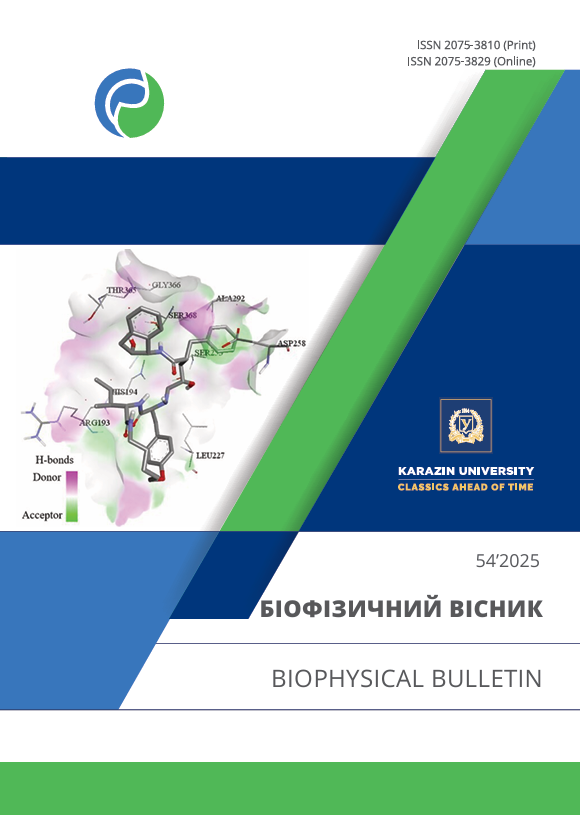
Results. The binding affinity of the studied ligands with the S protein-furin protease complexes was verified by molecular docking studies. PI8, nelfinavir, and remdesivir showed high binding affinity with the 7VHJ-5JXG structure due to the presence of amino acid residues at the furin cleavage site. The best docking scores of PI8 with 6VYB-5JXG, 6VXX-5JXG, and 7VHJ-5JXG complexes were -9.7 kcal/mol, -9.5 kcal/mol, and -9.9 kcal/mol, respectively. The interaction between the S protein-furin complexes and PI8 involves specific amino acid residues, primarily within the catalytic site of furin and the reactive site loop of PI8. Docking studies showed that remdesivir acts directly on the furin cleavage site of the S protein (in the 7VHJ-5JXG complex), forming energetically favorable interactions through hydrogen bonds and hydrophobic contacts, with a high binding affinity (binding energy score is -9.1 kcal/mol). The energetically favorable interactions of the 6VYB-5JXG, 6VXX-5JXG, and 7VHJ-5JXG complexes with nelfinavir are also confirmed by their low binding energy scores of -8.2 kcal/mol, -8.9 kcal/mol, and -9.3 kcal/mol, respectively.
Conclusion. According to the results of molecular docking, PI8, nelfinavir, and remdesivir demonstrate energetically favorable interactions with the studied complexes and can be considered promising inhibitors targeting the SARS-CoV-2 S protein–furin protease complexes.
Downloads
References
WHO. 2020. Coronavirus disease 2019 (COVID-19) weekly epidemiological updates. Available from: https://www.who.int/emergencies/diseases/novel-coronavirus-2019/situation-reports
Sanche S, Lin YT, Xu C, Romero-Severson E, Hengartner N, Ke R. High contagiousness and rapid spread of severe acute respiratory syndrome coronavirus 2. Emerg Infect Dis. 2020;26(7):1470–77. https://doi.org/10.3201/eid2607.200282
Sanders JM, Monogue ML, Jodlowski TZ, Cutrell JB. Pharmacologic treatments for coronavirus disease 2019 (COVID-19): A Review. JAMA. 2020;323(18):1824–36. https://doi.org/10.1001/jama.2020.6019
Khmil NV, Kolesnikov VG, Boiechko-Nemovcha AO. Binding characteristics of systemic glucocorticoids to the SARS-CoV-2 spike glycoprotein: in silico evaluation. Low Temp Phys. 2025;51:96–103 https://doi.org/10.1063/10.0034652
Zhang L, Lin D, Sun X, Curth U, Drosten C, Sauerhering L, et al. Crystal structure of SARS-CoV-2 main protease provides a basis for design of improved α-ketoamide inhibitors. Science. 2020;368(6489):409-12. https://doi.org/10.1126/science.abb3405
Khmil NV, Shestopalova AV, Kolesnikov VG, Boiechko-Nemovcha AO. Identification of potential corticosteroid binding sites on the SARS CoV-2 main protease Mpro- in silico docking study. Biophysical Bulletin. 2024;51:53–63. https://doi.org/10.26565/2075-3810-2024-51-04
Huang Y, Yang C, Xu XF, Xu W, Liu SW. Structural and functional properties of SARS-CoV-2 spike protein: potential antivirus drug development for COVID-19. Acta Pharmacol Sin. 2020;41(9):1141–49. https://doi.org/10.1038/s41401-020-0485-4
Zhang J, Xiao T, Cai Y, Chen B. Structure of SARS-CoV-2 spike protein. Curr Opin Virol. 2021;50:173–82. https://doi.org/10.1016/j.coviro.2021.08.010
Hulswit RJ, de Haan CA, Bosch BJ. Coronavirus spike protein and tropism changes. Adv Virus Res. 2016;96:29–57. https://doi.org/10.1016/bs.aivir.2016.08.004
Cai Y, Zhang J, Xiao T, Peng H, Sterling SM, Walsh RM, et al. Distinct conformational states of SARS-CoV-2 spike protein. Science. 2020;369(6511):1586–92. https://doi.org/10.1126/science.abd4251
Gur M, Taka E, Yilmaz SZ, Kilinc C, Aktas U, Golcuk M. Conformational transition of SARS-CoV-2 spike glycoprotein between its closed and open states. J Chem Phys. 2020;153(7):075101. https://doi.org/10.1063/5.0011141
Hoffmann M, Kleine-Weber H, Pöhlmann S. A Multibasic cleavage site in the spike protein of SARS-CoV-2 is essential for Infection of human lung cells. Mol Cell. 2020;78(4):779-84.e5. https://doi.org/10.1016/j.molcel.2020.04.022
Strobelt R, Adler J, Shaul Y. The Transmembrane protease serine 2 (TMPRSS2) non-protease domains regulating severe acute respiratory syndrome coronavirus 2 (SARS-CoV-2) spike-mediated virus entry. Viruses. 2023;15(10):2124. https://doi.org/10.3390/v15102124
Marcink TC, Kicmal T, Armbruster E, Zhang Z, Zipursky G, Golub KL, et al. Intermediates in SARS-CoV-2 spike-mediated cell entry. Sci Adv. 2022;8(33):eabo3153. https://doi.org/10.1126/sciadv.abo3153
Peacock TP, Goldhill DH, Zhou J, Baillon L, Frise R, Swann OC, et al. The furin cleavage site in the SARS-CoV-2 spike protein is required for transmission in ferrets. Nat Microbiol. 2021;6(7):899–909. https://doi.org/10.1038/s41564-021-00908-w
Coutard B, Valle C, de Lamballerie X, Canard B, Seidah NG, Decroly E. The spike glycoprotein of the new coronavirus 2019-nCoV contains a furin-like cleavage site absent in CoV of the same clade. Antiviral Res. 2020;176:104742. https://doi.org/10.1016/j.antiviral.2020.104742
Holmes EC, Goldstein SA, Rasmussen AL, Robertson DL, Crits-Christoph A, Wertheim JO, et al. The origins of SARS-CoV-2: A critical review. Cell. 2021;184(19):4848–56. https://doi.org/10.1016/j.cell.2021.08.017
Cheng YW, Chao TL, Li CL, Chiu MF, Kao HC, Wang SH, et al. Furin inhibitors block SARS-CoV-2 spike protein cleavage to suppress virus production and cytopathic effects. Cell Rep. 2020;33(2):108254. https://doi.org/10.1016/j.celrep.2020.108254
Brown AJ, Won JJ, Graham RL, Dinnon KH, Sims AC, Feng JY, et al. Broad spectrum antiviral remdesivir inhibits human endemic and zoonotic deltacoronaviruses with a highly divergent RNA dependent RNA polymerase. Antiviral Res. 2019;169:104541. https://doi.org/10.1016/j.antiviral.2019.104541
Nguyen TH, Guedj J, Anglaret X, Laouénan C, Madelain V, Taburet AM, et al. Favipiravir pharmacokinetics in Ebola-Infected patients of the JIKI trial reveals concentrations lower than targeted. PLoS Negl Trop Dis. 2017;11(2):e0005389. https://doi.org/10.1371/journal.pntd.0005389
Garriga C, Pérez-Elías MJ, Delgado R, Ruiz L, Nájera R, Pumarola T, et al. Mutational patterns and correlated amino acid substitutions in the HIV-1 protease after virological failure to nelfinavir- and lopinavir/ritonavir-based treatments. J Med Virol. 2007;79(11):1617–28. https://doi.org/10.1002/jmv.20986
Agostini ML, Andres EL, Sims AC, Graham RL, Sheahan TP, Lu X, et al. Coronavirus susceptibility to the antiviral remdesivir (GS-5734) is mediated by the viral polymerase and the proofreading exoribonuclease. mBio. 2018;9(2):e00221-18. https://doi.org/10.1128/mBio.00221-18
Grein J, Ohmagari N, Shin D, Diaz G, Asperges E, Castagna A, et al. Compassionate use of remdesivir for patients with severe Covid-19. N Engl J Med. 2020;382(24):2327–36. https://doi.org/10.1056/NEJMoa2007016
Driouich JS, Cochin M, Lingas G, Moureau G, Touret F, Petit PR, et al. Favipiravir antiviral efficacy against SARS-CoV-2 in a hamster model. Nat Commun. 2021;12(1):1735. https://doi.org/10.1038/s41467-021-21992-w
Jia Y, Tian W, Li Y, Teng Y, Liu X, Li Z, et al. Chloroquine: Rapidly withdrawing from first-line treatment of COVID-19. Heliyon. 2024;10(17):e37098. https://doi.org/10.1016/j.heliyon.2024.e37098
Søndergaard CR, Olsson MH, Rostkowski M, Jensen JH. Improved treatment of ligands and coupling effects in empirical calculation and rationalization of pKa values. J Chem Theory Comput. 2011;7(7):2284–95. https://doi.org/10.1021/ct200133y
Trott O, Olson AJ. AutoDock Vina: improving the speed and accuracy of docking with a new scoring function, efficient optimization, and multithreading. J Comput Chem. 2010;31(2):455–61. http://doi.org/10.1002/jcc.21334
O'Boyle NM, Banck M, James CA, Morley C, Vandermeersch T, Hutchison GR. Open Babel: An open chemical toolbox. J Cheminform. 2011;3:33. https://doi.org/10.1186/1758-2946-3-33
Dahms SO, Jiao GS, Than ME. Structural studies revealed active site distortions of human furin by a small molecule inhibitor. ACS Chem Biol. 2017;12(5):1211–16. https://doi.org/10.1021/acschembio.6b01110
Kozakov D, Brenke R, Comeau SR, Vajda S. PIPER: an FFT-based protein docking program with pairwise potentials. Proteins. 2006;65(2):392–406. https://doi.org/10.1002/prot.21117
Bashir A, Li S, Ye Y, Zheng Q, Knanghat R, Bashir F, et al. SARS-CoV-2 S protein harbors furin cleavage site located in a short loop between antiparallel β-strand. Int J Biol Macromol. 2024;281(Pt 1):136020. https://doi.org/10.1016/j.ijbiomac.2024.136020
Vankadari N. Structure of furin protease binding to SARS-CoV-2 spike glycoprotein and implications for potential targets and virulence. J Phys Chem Lett. 2020;11(16):6655–63. https://doi.org/10.1021/acs.jpclett.0c01698
Bollavaram K, Leeman TH, Lee MW, Kulkarni A, Upshaw SG, Yang J, et al. Multiple sites on SARS-CoV-2 spike protein are susceptible to proteolysis by cathepsins B, K, L, S, and V. Protein Sci. 2021;30(6):1131–43. https://doi.org/10.1002/pro.4073
Bosch BJ, Bartelink W, Rottier PJ. Cathepsin L functionally cleaves the severe acute respiratory syndrome coronavirus class I fusion protein upstream of rather than adjacent to the fusion peptide. J Virol. 2008;82(17):8887–90. https://doi.org/10.1128/JVI.00415-08
Van Lam van T, Ivanova T, Hardes K, Heindl MR, Morty RE, Böttcher-Friebertshäuser E, et al. Design, synthesis, and characterization of macrocyclic inhibitors of the proprotein convertase furin. ChemMedChem. 2019;14(6):673–85. https://doi.org/10.1002/cmdc.201800807
Gordon CJ, Tchesnokov EP, Woolner E, Perry JK, Feng JY, Porter DP, et al. Remdesivir is a direct-acting antiviral that inhibits RNA-dependent RNA polymerase from severe acute respiratory syndrome coronavirus 2 with high potency. J Biol Chem. 2020;295(20):6785–97. https://doi.org/10.1074/jbc.RA120.013679
Nguyen HL, Thai NQ, Truong DT, Li MS. Remdesivir strongly binds to both RNA-dependent RNA polymerase and main protease of SARS-CoV-2: evidence from molecular simulations. J Phys Chem B. 2020;124(50):11337–48. https://doi.org/10.1021/acs.jpcb.0c07312
Eweas AF, Alhossary AA, Abdel-Moneim AS. Molecular docking reveals ivermectin and remdesivir as potential repurposed drugs against SARS-CoV-2. Front Microbiol. 2021;11:592908. https://doi.org/10.3389/fmicb.2020.592908
Sukeishi A, Itohara K, Yonezawa A, Sato Y, Matsumura K, Katada Y, et al. Population pharmacokinetic modeling of GS-441524, the active metabolite of remdesivir, in Japanese COVID-19 patients with renal dysfunction. CPT: Pharmacomet Syst Pharmacol. 2022;11(1):94–103. https://doi.org/10.1002/psp4.12736
Thomas G. Furin at the cutting edge: from protein traffic to embryogenesis and disease. Mol Cell biol. 2002;3(10):753–66. https://doi.org/10.1038/nrm934
Jorge A, Ung C, Young LH, Melles RB, Choi HK. Hydroxychloroquine retinopathy - implications of research advances for rheumatology care. Nat Rev Rheumatol. 2018;14(12):693–703. https://doi.org/10.1038/s41584-018-0111-8
Badraoui R, Adnan M, Bardakci F, Alreshidi MM. Chloroquine and hydroxychloroquine interact differently with ACE2 domains reported to bind with the coronavirus spike protein: mediation by ACE2 polymorphism. Molecules. 2021;26(3):673. https://doi.org/10.3390/molecules26030673
El Khatabi K, Aanouz I, Alaqarbeh M, Ajana MA, Lakhlifi T, Bouachrine M. Molecular docking, molecular dynamics simulation, and ADMET analysis of levamisole derivatives against the SARS-CoV-2 main protease (MPro). Bioimpacts. 2022;12(2):107–13. https://doi.org/10.34172/bi.2021.22143
Shannon A, Selisko B, Le NT, Huchting J, Touret F, Piorkowski G, et al. Rapid incorporation of favipiravir by the fast and permissive viral RNA polymerase complex results in SARS-CoV-2 lethal mutagenesis. Nat Commun. 2020;11(1):4682. https://doi.org/10.1038/s41467-020-18463-z
Yadav P, Rana M, Chowdhury P. DFT and MD simulation investigation of favipiravir as an emerging antiviral option against viral protease (3CLpro) of SARS-CoV-2. J Mol Struct. 2021;1246:131253. https://doi.org/10.1016/j.molstruc.2021.131253
Citations
Effect of antivirals and a peptidomimetic inhibitor on furin conformational stability: Molecular docking and molecular dynamics simulations
Khmil N. V. & Shestopalova A. V. (2026) Biopolymers and Cell
Crossref
Copyright (c) 2025 N. V. Khmil, A. V. Shestopalova, V. G. Kolesnikov

This work is licensed under a Creative Commons Attribution 4.0 International License.
Authors who publish with this journal agree to the following terms:
- Authors retain copyright and grant the journal right of first publication with the work simultaneously licensed under a Creative Commons Attribution License that allows others to share the work with an acknowledgement of the work's authorship and initial publication in this journal.
- Authors are able to enter into separate, additional contractual arrangements for the non-exclusive distribution of the journal's published version of the work (e.g., post it to an institutional repository or publish it in a book), with an acknowledgement of its initial publication in this journal.
- Authors are permitted and encouraged to post their work online (e.g., in institutional repositories or on their website) prior to and during the submission process, as it can lead to productive exchanges, as well as earlier and greater citation of published work (See The Effect of Open Access).


