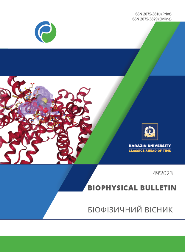
Молекулярний докінг сироваткового альбуміну людини з детермінантами пеніциліну G
Анотація
Актуальність. Сироватковий альбумін людини (САЛ) є основним фармакокінетичним ефектором багатьох ліків, в тому числі пеніциліну G та його метаболітів. Гострою проблемою практичної медицини є реакції гіперчутливості негайного типу, які зумовлені токсичністю пеніцилінів (близько 8% проти інших препаратів), що супроводжуються патологією шкіри, анафілаксією та летальністю.
Мета роботи. Метою цього дослідження є опис структур комплексів САЛ-детермінанти пеніциліну G і виявлення сприятливих сайтів зв'язування та амінокислотних залишків, які залучені до взаємодії.
Матеріали та методи. Кристалічна структура САЛ (ID:1AO6 з Protein Data Bank) (www.rcsb.org) була вибрана як мішень для докінгу. Для отримання уявлення про взаємодію САЛ з основними (бензіл пеніцилоїл G, пеніциланова кислота) і другорядними (пеніциламін, пеніцилоєва кислота, пенілоєва кислота) детермінантами пеніциліну G, були застосовані методи молекулярного докінгу (AutoDock Tools 1.5.7, AutoDock Vina 1.1.2). Візуалізація результатів докінгу була реалізована в PyMol 2.5. Для оцінки потенційних сайтів зв’язування був використаний Protein Plus сервер (https://proteins.plus). Для ідентифікації нековалентних взаємодій між САЛ та його лігандами був застосований засіб PLIP (https://plip-tool.biotec.tu-dresden.de).
Результати. Дані молекулярного моделювання свідчать, що основні детермінанти пеніциліну G беруть участь в утворенні водневих зв’язків з такими залишками САЛ, як Trp214, Arg218, His242 та Asn295; для другорядних детермінант — Asp108, His146, Tyr148, Ser193, Arg197, Gln204. Обидва типи детермінант розташовуються в гідрофобній порожнині субдоменів IIA та IB. Гідрофобні взаємодії присутні переважно між детермінантами пеніциліну G і амінокислотними залишками субдомену IIIA, такими як Ala350, Asp451, Tyr452 і Gln459.
Висновки. Вивчення комплексів САЛ-детермінанти пеніциліну G має важливе значення в патогенезі алергії на антибіотики. Виявлення специфічних сайтів зв’язування може бути корисним для розробки та синтезу нових імуногенних антигенів (комплексів основних і другорядних детермінант пеніциліну G із САЛ), які зможуть стимулювати імунну систему та виробляти специфічні антитіла для запобігання алергічної реакції.
Завантаження
Посилання
Batra A, Roemhild R, Rousseau E, Franzenburg S, Niemann S, Schulenburg H. High potency of sequential therapy with only β-lactam antibiotics. eLife. 2021;10:e68876. https://doi.org/10.7554/eLife.68876
Turner J, Muraoka A, Bedenbaugh M, Childress B, Pernot L, Wiencek M, et al. The chemical relationship among beta-lactam antibiotics and potential impacts on reactivity and decomposition. Front Microbiol. 2022;13:807955. https://doi.org/10.3389/fmicb.2022.807955
Maguire M, Hayes BD, Fuh L. Beta-lactam antibiotic test doses in the emergency department. World Allergy Organ J. 2020;13(1):100093. https://doi.org/10.1016/j.waojou.2019.100093
Brockow K. Drug Allergy: Definitions and Phenotypes. In: Khan DA, Banerji A, editors. Drug Allergy Testing. Elsevier; 2018. p. 19–26. https://doi.org/10.1016/B978-0-323-48551-7.00003-1
Wilkerson GR. Drug Hypersensitivity Reactions. Emerg Med Clin N Am. 2022;40:39–55. https://doi.org/10.1016/j.emc.2021.09.001
Canzani D, Aldeek F. Penicillin G’s function, metabolites, allergy, and resistance. J Nutr Hum Health. 2017;1(1):28–40. http://doi.org/10.35841/nutrition-human-health.1.1.28-40
Fanali G, di Masi A, Trezza V, Marino M, Fasano M, Ascenzi P. Human serum albumin: from bench to bedside. Mol Aspects Med. 2012;33(3):209–90. https://doi.org/10.1016/j.mam.2011.12.002
Yamasaki K, Chuang VT, Maruyama T, Otagiri M. Albumin-drug interaction and its clinical implication. Biochim Biophys Acta. 2013;1830(12):5435–43. https://doi.org/10.1016/j.bbagen.2013.05.005
He XM, Carter DC. Atomic structure and chemistry of human serum albumin. Nature. 1992;358(6383):209–15. https://doi.org/10.1038/358209a0
Sugio S, Kashima A, Mochizuki S, Noda M, Kobayashi K. Crystal structure of human serum albumin at 2.5 Å resolution. Protein Eng. 1999;12: 439–46. https://doi.org/10.1093/protein/12.6.439
Vallianatou T, Lambrinidis G, Tsantili-Kakoulidou A. In silico prediction of human serum albumin binding for drug leads. Expert Opin. Drug. Discov. 2013;8(5):583–95. https://doi.org/10.1517/17460441.2013.777424
Yang F, Zhang Y, Liang H. Interactive association of drugs binding to human serum albumin. Int J Mol Sci. 2014;15(3):3580–95. http://doi.org/10.3390/ijms15033580
Calderaro A, Maugeri A, Magazù S, Laganà G, Navarra M, Barreca D. Molecular basis of interactions between the antibiotic nitrofurantoin and human serum albumin: a mechanism for the rapid drug blood transportation. Int J Mol Sci. 2021;22(16):8740. https://doi.org/10.3390/ijms22168740
Abou-Zied OK, Al-Shihi OI. Characterization of subdomain IIA binding site of human serum albumin in its native, unfolded, and refolded states using small molecular probes. J Am Chem Soc. 2008;130:10793–801. https://doi.org/10.1021/ja8031289
Seedher N, Agarwal P. Interaction of some isoxazolyl penicillins with human serum albumin. J Biol Sci. 2006;6(1):167–72. http://doi.org/10.3923/jbs.2006.167.172
Ahmad B, Parveen S, Khan RH. Effect of albumin conformation on the binding of ciprofloxacin to human serum albumin: a novel approach directly assigning binding site. Biomacromolecules. 2006;7:1350–56. http://doi.org/10.1021/bm050996b
Barabosa S, Taboada P, Attwood D, Mosquera V. Thermodynamic properties of the complex formed by interaction of two anionic amphiphilic penicillins with human serum albumin. Langmuir. 2003;19:10200–204. https://doi.org/10.1021/la035106x
DiPiro JT, Adkinson NF, Hamilton RG. Facilitation of penicillin haptenation to serum proteins. Antimicrob Agents Chemother. 1993;37(7):1463–67. https://doi.org/10.1128/AAC.37.7.1463
Blanca M, Mayorga C, Perez E, Suau R, Juarez C, Vega JM, et al. Determination of IgE antibodies to the benzyl penicilloyl determinant. A comparison between poly-L-lysine and human serum albumin as carriers. J Immunol Methods. 1992;153(1-2):99–105. https://doi.org/10.1016/0022-1759(92)90311-g
Zhao Z, Batley M, D'Ambrosio C, Baldo BA. In vitro reactivity of penicilloyl and penicillanyl albumin and polylysine conjugates with IgE-antibody. J Immunol Methods. 2000;242(1–2):43–51. https://doi.org/10.1016/s0022-1759(00)00213-1
Trott O, Olson AJ. AutoDock Vina: improving the speed and accuracy of docking with a new scoring function, efficient optimization, and multithreading. J Сomp chem. 2010;31(2):455–61. http://doi.org/10.1002/jcc.21334
Morris GM, Huey R, Olson AJ. Using AutoDock for ligand-receptor docking. Current Protocols in Bioinformatics. 2008;24(1);8141–440. http://doi.org/10.1002/0471250953.bio0814s24
Søndergaard CR, Olsson MH, Rostkowski M, Jensen JH. Improved treatment of ligands and coupling effects in empirical calculation and rationalization of pKa values. J Chem Theory Comput. 2011;7(7):2284–95. https://doi.org/10.1021/ct200133y
Schrödinger L, DeLano W. PyMOL [Internet]. [cited 2020]. Available from: http://www.pymol.org/pymol
Volkamer A, Griewel A, Grombacher T, Rarey M. Analyzing the topology of active sites: on the prediction of pockets and subpockets. J Chem Inf Model. 2010;50(11):2041–52. https://doi.org/10.1021/ci100241y
Volkamer A, Kuhn D, Grombacher T, Rippmann F, Rarey M. Combining global and local measures for structure-based druggability predictions. J Chem Inf Model. 2012;52(2):360–72. https://doi.org/10.1021/ci200454v
Stierand K, Maass PC, Rarey M. Molecular complexes at a glance: automated generation of two-dimensional complex diagrams. Bioinformatics. 2006;22(14):1710–16. https://doi.org/10.1093/bioinformatics/btl150
Fricker PC, Gastreich M, Rarey M. Automated drawing of structural molecular formulas under constraints. J Chem Inf Comput Sci. 2004;44(3): 1065–78. https://doi.org/10.1021/ci049958u
Adasme MF. PLIP 2021: expanding the scope of the protein-ligand interaction profiler to DNA and RNA. Nucl Acids Res. 2021;49(1): 530–34. http://doi.org/10.1093/nar/gkab294
Retnaningtyas E, Sumitro SB, Soeatmadji DW, Widjayanto E. Molecular dynamics simulation for revealing the role of water molecules on conformational change of human serum albumin. Int J Pharm Clin Res. 2016;8(3):158–61. http://impactfactor.org/PDF/IJPCR/8/IJPCR,Vol8,Issue3,Article1.pdf
Keswani N, Choudhary S, Kishore N. Interaction of weakly bound antibiotics neomycin and lincomycin with bovine and human serum albumin: biophysical approach. J Biochem. 2010;148(1):71–84. http://doi.org/10.1093/jb/mvq035
Li Q, Zhang T, Bian L. Recognition and binding of β-lactam antibiotics to bovine serum albumin by frontal affinity chromatography in combination with spectroscopy and molecular docking. J Chromatogr B: Anal Technol Biomed Life Sci. 2016;1014:90–101. https://doi.org/10.1016/j.jchromb.2016.02.005
Yvon M, Anglade P, Wal JM. Binding of benzyl penicilloyl to human serum albumin. Evidence for a highly reactive region at the junction of domains 1 and 2 of the albumin molecule. FEBS Lett. 1989;247(2):273–78. https://doi.org/10.1016/0014-5793(89)81351-1
Zhang Y, Cao Y, Li Y, Zhang X. Interactions between human serum albumin and sulfadimethoxine determined using spectroscopy and molecular docking. Molecules. 2022;27:1526. https://doi.org/10.3390/molecules27051526
Цитування
DHEA-carbamate derivatives as dual cholinesterase inhibitors: Integration of enzymatic and biomolecular interactions in Alzheimer's disease
Nar Kubra, Erdagi Sevinc Ilkar & Ozbagci Duygu Inci (2025) Bioorganic Chemistry
Crossref
Binding characteristics of systemic glucocorticoids to the SARS-CoV-2 spike glycoprotein: In silico evaluation
Khmil N. V., Kolesnikov V. G. & Boiechko-Nemovcha A. O. (2025) Low Temperature Physics
Crossref
Автори, які публікуються у цьому журналі, погоджуються з наступними умовами:
- Автори залишають за собою право на авторство своєї роботи та передають журналу право першої публікації цієї роботи на умовах ліцензії Creative Commons Attribution License, котра дозволяє іншим особам вільно розповсюджувати опубліковану роботу з обов'язковим посиланням на авторів оригінальної роботи та першу публікацію роботи у цьому журналі.
- Автори мають право укладати самостійні додаткові угоди щодо неексклюзивного розповсюдження роботи у тому вигляді, в якому вона була опублікована цим журналом (наприклад, розміщувати роботу в електронному сховищі установи або публікувати у складі монографії), за умови збереження посилання на першу публікацію роботи у цьому журналі.
- Політика журналу дозволяє і заохочує розміщення авторами в мережі Інтернет (наприклад, у сховищах установ або на особистих веб-сайтах) рукопису роботи, як до подання цього рукопису до редакції, так і під час його редакційного опрацювання, оскільки це сприяє виникненню продуктивної наукової дискусії та позитивно позначається на оперативності та динаміці цитування опублікованої роботи (див. The Effect of Open Access).


